
I took a trip out to University of Pennsylvania Hospital today with my CT Scan and MRI results in tow to start the process to have my right ear implanted. My right ear was a candidate last year but I still have to go through the whole process again minus all the tests. I don't have to redo the CT Scan, MRI or the balance tests. I don't need to have any audiological testing done because it's not as though my ears have gotten better :) In fact, my right ear, the one I wear a hearing aid in has finally gone kaput. I might as well should put it to good use! I am still a candidate and I don't have a surgery date yet because there is an issue with my oh-so-wonderful-union-backed-health-insurance canceling the contract with the hospital and they want to assure they get approval so I am not stuck with a hundred twenty-thousand dollar bill. And quite frankly, I want assurance that I am not going to get stuck with a hundred twenty-thousand dollar bill too. But that is another blog entirely.
When I saw the doctor, he came in and sat on the chair. He leaned back and crossed his legs, interlocked his fingers and rested them gingerly on his lap while he gave me the best present that I could have asked for: the answer to the age-old question of why I am deaf since no one seems to know why I have a hearing loss since I am the only one on both sides of the family that is deaf.
"Abbie, you have what is called Enlarged Vestibular Aqueduct." He says.
"En-larged Ves-ti-bul-ar Aq-ue-duct." I repeated after him, syllable by syllable.
I presumed to steam roll him with questions. With the recent advancement of MRI technology, it has made it much easier to diagnosis this. A CT Scan can appear normal but with an MRI you can actually see the enlarged duct and sac. He explained to me that I was born with it. Most children that are born with this don't lose their hearing until three or four years old since this is when the Vestibular Aqueduct reaches its normal adult size. Figures, I would have something enlarged in my ears. My ass is enlarged. My chest is enlarged. So why not my something in my ears! The second I left, I was determined to become a self proclaimed expert via the way of the Blackberry of this Enlarged Vestibular Aqueduct Syndrome. And everything that I have read so far, fits perfectly. From the reason I feel as if I have fluid in my ears after I get my head jarred to the sudden deafness to the progressive hearing loss and how the one side worse than the other. It fits. This will be a separate blog too. I know, I know, what is the point of this blog you ask? Just hold on to your candy canes, I'm getting there.
My answer to my deafness has long be undetermined but I finally have an answer or so I thought.
Since I was stuck in traffic for a greater part of the day, I spent much of my time on my blackberry reading about this Enlarged Vestibular Aqueduct Syndrome. This shouldn't surprise you because when I get interested something, I research it to the death. But anyway, I finally got home from finishing up some Christmas shopping. I decided in all my professional incapacity to take a look at my MRI results. I never took a look at them before so I figured why the heck not!
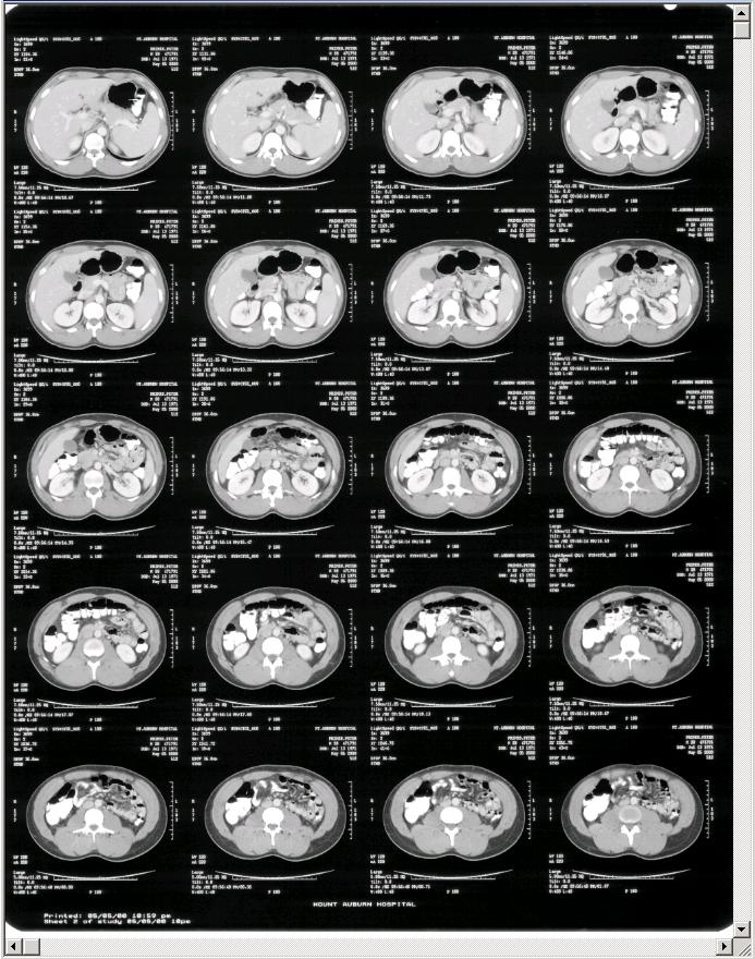
I cracked open the huge Manila envelope and as I removed the MRI sheets as if they were made of glass. There was a ton of them! I carefully pick one up and hold one up to the light and started to admire each of the images on the film. I couldn't help but think they look like a very boring black and white Andy Warhol painting. They were arranged four across and five down: 20 images total on a single sheet of film. Like this:
I had no idea what I was looking at but I just so happened to take notice of a name at the upper right corner of each tile - a Wayne Something. Note: I changed the name to respect this person privacy.
First I thought it was the name of the person who performed the test or the name of the radiologists but I read further: Pat.: Wayne Something born in 1952.
Ok. That means patient.
I picked up another sheet of film. Wayne Something.
I picked up another one and sure enough, Wayne Something again!
And another one, Wayne Something.
I'm sensing a pattern here.
Yet another sheet, WAYNE SOMETHING
Picked up another sheet and saw a familiar name, MINE!
All in all, a total of four sheets belong to me.
All twenty other sheets belonged to Wayne Something born in 1952!
Ok. I thought maybe the hospital might have incidentally given me back someone else films but I checked out the date that Wayne Something born in 1952 had his MRI done. It matched the same date that I had mine done.
I’m attempting to think logically here because I am so frigging furious and I am pms'ing and all the dark chocolate in the world isn't calming me down. The answer that my entire family and I have all been waiting for was just handed to me on a silver platter and NOW, the possibility that the diagnosis was based on WaynE Something born in 1952 films and not my four friggen MRI films.
DO I LOOK LIKE A FIFTY YEAR OLD MAN TO ANY OF YOU?!
No, I didn't think so either. My so-called logical thinking has lead me to conclude that when I went to pick up my MRI films, they gave me Wayne's films and I never had a full set of MRI films to begin with.
What is really upsetting me is what if the doctor based his diagnosis of having Enlarged Vestibular Aqueduct Syndrome on HIS sheets and over looked the name?
That means I am back to square one without an answer to why I am deaf.
The hell I'm going back to square one without a fight. First thing tomorrow morning, I'm calling the MRI place, calmly, and tell them what happened and politely request (demand) that I get my full copy of my MRI results. I am sincerely hoping that they still have a copy of my MRI films because this is dating as back to early 2007. I will request the radiologist to measure the size of my vestibular aqueducts to see whether the doctor diagnosis is correct.
I practically screamed my friends ear off on the phone tonight reciting this entire SCREW UP to her and I decided it was time to give her a word in edgewise because I puffed out all the oxygen in my lungs. First thing she does, is her worst impression of Barry White singing,
"Happy Holidays!"
Stay tuned folks and have a great holiday! :)
Tuesday, December 23, 2008
Finally, an Answer to Why I'm Deaf or a Total Medical Screw Up!?
Wednesday, December 03, 2008
MRIs and Cochlear Implants
Let's talk MRIs.
When I went to Chicago for the ALDA convention, I met several people that didn't want to get a cochlear implant because they need an MRI every six months. I will admit when I first started researching cochlear implants, MRIs was not a major concern of mine. I just read the I can have a MRI done if the magnet was removed. Fine. Great! That is all I needed to know but now I realize how much it means to others that suffer from other illnesses where they require MRIs.
Why would a person need to get one? MRIs provide better contrast in soft tissue, which helps to distinguish between normal and diseased tissue. MRIs do not show bones like a CAT scan or X-Ray. Brain tumors, strokes, multiple sclerosis and Neurofibromatosis, type 2 (NF2), are diagnosed by an MRI. Which means anything metal - paper clips, pens, keys, jewelry, scissors, underwire in your bra, belts, glasses and any other small objects can be pulled out of pockets and off the body or out of the body can become dangerous projectiles hurdling at the opening of the tube at incredibly high speeds.
Joy. Its a good thing that they make you remove anything metal.
Could you imagine if someone left a tongue piercing in and they turned the MRI machine on? Ouch.
Anyway, I did me a little research on MRIs. The magnet in an MRI system is rated using a unit of measure known as a Tesla and they are grouped into three fields.
Low-Field = Under .2 Tesla
Mid-Field = .2 to 0.6 Tesla
High-Field = 1.0 to 1.5 Tesla
What is the difference between low-field and high-field? The high-field setup has superior image quality AND has a higher rate of detecting tumor remnants. This abstract that I found supports that statement. The next generation of MRIs are circulating around at the strength of 3.0 Tesla.
Sounds like the higher the Tesla - the better the detection rate. I would imagine it would be like going from a two mega pixel camera to a ten mega pixel camera.
Now both Advanced Bionics, HiResolution Bionic Ear System's HiRes 90K implant and Cochlear Americas, Nucleus Freedom is MRI Safe up to 1.5 Tesla with the internal magnet removed.
I took a look at Med-El's website and discovered in bold letters, MRI Safe - Without Magnet Removal. Leaping lizards, no faking! They don't require the internal magnet to be removed. In fact, it is designed where the magnet can't be removed at all.
That cool!
But then I read the fine print:

In the US, PULSARCI100 and SONATATI100 are currently approved for use at a scanner strength of 0.2 Tesla.
Oh. That means recipients of a Med-El device can only use MRI's rated at low-field strength of 0.2 Tesla where they could be sacrificing image quality that could lead to a potential misdiagnoses.
What if one with a Med-EL device wants a high-field MRI that has a better image quality and higher rate of detection? Does that mean the entire implant has to be removed because they don't have a removable magnet?
Yikes.
This is a link to an article that talks about the latest MRI machines that are rated 3.0 Tesla which can demagnetize an implant. It also discusses how there is permanent damage to devices with non-removable magnets such as Med-El's PULSARCI100 and SONATATI100.
However, I'm privileged to know someone in my harem of cochlear implant users that had an MRI done and had the internal magnet removed and what he had to say really calmed my nerves if I ever had to get one.
First of all, it seems to be kind of a rare event. My surgeon has performed over 550 implant surgeries and has never had to do this procedure (taking the internal magnets out, then reinserting new magnets). In fact, of the 700+ Midwest Ear Institute patients, I believe I am the first to have an MRI. To get an MRI, the internal magnets need to be removed from the implant, then you get in the tube, then back to the OR to have new, sterile magnets and stitched up. The thought of having the internal magnets taken out probably bothers some people but it shouldn't - it was not a big deal at all. In fact, they asked if I wanted to be sedated and I said no - so they just did a local and it was fine. There is a little pain...but very little, and easily handled with OTC pain relievers. In fact, I haven't needed any today at all. I was going to write a great, detailed account of this but it is such a non-event that there is little to write. Kind of like getting some stitches in your head - that's it. The most irritating thing is being inside the MRI tube - at least you can't hear it though, because you are completely deaf while inside.
I wouldn't go get an MRI for fun, but if it is suggested that you need one, please do not hesitate to do it. An MRI is an incredible piece of technology and can be a difference maker in terms of diagnosing certain things.
And that is all he said folks!


